
Diabetic Retinopathy: Why Eye Damage from Diabetes Shows No Symptoms Until Vision Is Already Gone


The Eye Bleeds Before You Know ItDiabetes does not attack the eye the way an infection does, with pain, redness, and urgency. It works on the tiny blood vessels at the back of the eye, the retina, over months and years. High blood sugar makes those vessel walls weak and leaky. They bulge into microaneurysms, seep fluid into retinal tissue, and eventually rupture. Through all of this, the person sees nothing unusual. The retina has no pain receptors. There is no signal to send.
 A 2019 study published in the Indian Journal of Ophthalmology estimated that roughly 18 percent of people with diabetes in India have some form of retinopathy, and a significant proportion of them are unaware of it. The retina can lose a substantial portion of its functioning cells before central vision noticeably dims. By the time a patient notices blurring, the peripheral retinal damage is often already severe.
Why the Brain Fills In the GapsThe visual system is designed to compensate. When small patches of the retina stop working, the brain interpolates, it fills in the missing information using surrounding signals, the way it fills in the blind spot every human eye has at the optic nerve. This is not a flaw. It is a feature that becomes a liability in retinopathy. The brain masks early damage so effectively that patients report normal vision even when retinal imaging shows extensive lesions.
Proliferative diabetic retinopathy, the advanced stage, is where new abnormal blood vessels grow across the retina in an attempt to restore oxygen supply. These vessels are fragile and prone to large hemorrhages. When they bleed into the vitreous, the gel filling the eye, vision can go dark almost overnight. That sudden event feels like the beginning of the problem. It is the end of a long, silent progression.
The Blood Sugar and Retina Connection Is Not LinearOne of the most clinically important findings from the landmark UKPDS (United Kingdom Prospective Diabetes Study) is that retinopathy risk tracks with cumulative glycemic exposure, not just current HbA1c. A person who kept blood sugar poorly controlled for a decade and then brought it under control still carries elevated retinal risk. The damage already done to vessel walls does not fully reverse. This is why ophthalmologists speak of a metabolic memory, the retina remembers years of high glucose even after the numbers improve.
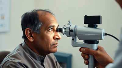
In practical terms, this means that a patient whose diabetes is now well-managed can still be developing retinopathy from earlier years of poor control. The absence of current symptoms says nothing about what is already under way in the retinal microvasculature. What Screening Actually CatchesA dilated fundus examination, where the pupil is widened with drops and the retina examined directly, can detect microaneurysms, hemorrhages, and new vessel growth long before any symptom appears. Optical coherence tomography (OCT) goes further, producing cross-sectional images of retinal layers that reveal fluid accumulation invisible to a standard exam.
The All India Ophthalmological Society recommends that every person diagnosed with type 2 diabetes get a dilated eye exam at diagnosis, and annually thereafter. For type 1 diabetes, the exam should begin within five years of diagnosis. In practice, large-scale surveys across Indian cities including Chennai and Hyderabad have found that fewer than 30 percent of diabetic patients report ever having had a dilated retinal exam. The gap between recommendation and reality is where blindness happens.
The Treatment Window That Closes QuietlyLaser photocoagulation, anti-VEGF injections, and vitrectomy surgery are all effective, but their effectiveness depends on timing. Anti-VEGF drugs, injected directly into the eye, can reduce macular edema and halt the growth of abnormal vessels. They work best when the macula, the central retina responsible for reading and face recognition, is still structurally intact. Once macular cells die, no injection restores them.
Why the Brain Fills In the GapsThe visual system is designed to compensate. When small patches of the retina stop working, the brain interpolates, it fills in the missing information using surrounding signals, the way it fills in the blind spot every human eye has at the optic nerve. This is not a flaw. It is a feature that becomes a liability in retinopathy. The brain masks early damage so effectively that patients report normal vision even when retinal imaging shows extensive lesions.
Next Story